
Mao Miyamoto
Tympanic cavity walls (English)
Tympanic cavity walls (English)
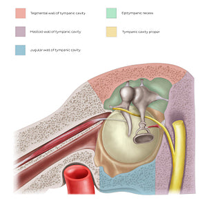
Sagittal section of the middle ear viewed from a medial perspective. The tympanic cavity has two main parts: the tympanic cavity proper and the epitympanic recess. The tympanic cavity proper is located medially to the tympanic membrane, while the epitympanic recess lies above the level of the tympanic membrane, next to the mastoid air cells. The tympanic cavity is shaped like a cube, containing 6 walls: membranous, tegmental, jugular, mastoid, labyrinthine and carotid wall, with the latter 2 not shown in this section. The membranous (lateral) wall is formed by the tympanic membrane and the squamous part of temporal bone. The labyrinthine (medial) wall separates the tympanic cavity from the labyrinth. The tegmental wall (roof) is a thin plate of bone that separates the tympanic cavity from the cranial cavity, while the jugular wall (floor) separates it from the jugular vein and the carotid artery below. The carotid (anterior) wall corresponds to the carotid canal and contains the tympanic opening of the auditory tube, while the mastoid (posterior) wall partly separates the tympanic cavity from the mastoid antrum.
Regular price
$7.56 USD
Regular price
Sale price
$7.56 USD
Unit price
per
Couldn't load pickup availability

#D9C435
#A88E5F
#A91D1E
#594136
#EC9291
#A4D0CF