
Paul Kim
Testis and epididymis (Latin)
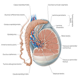
Testis and epididymis (Latin)
A section of the testis and surrounding connective tissue layers has been removed in order to visualize the internal structure of the testis. The tubuli seminiferi contorti form the bulk of the testis and converge to become the tubuli seminiferi recti as they approach the hilum, where they form the rete testis. The ductuli efferentes arise from the rete testis and converge to form the caput epididymidis and corpus epididymidis. The ductuli continue to unite within the epididymis, forming a single duct, known as the cauda epididymidis, which continues as the ductus deferens. The testis is enveloped by the tunica vaginalis and the tunica albuginea. The tunica vaginalis has two laminae: the lamina visceralis tunicae vaginalis testis and the lamina parietalis tunicae vaginalis testis between which is a potential space known as the cavitas tunicae vaginalis. The tunica albuginea envelopes only the testis, while the tunica vaginalis envelopes the testis and much of the epididymis.
Regular price
$7.56 USD
Regular price
Sale price
$7.56 USD
Unit price
per
Couldn't load pickup availability

#0574CB
#638FAC
#1667A6
#3B3D4A
#E7B29C
#CAB7AA