
Paul Kim
Testis and epididymis (English)
Testis and epididymis (English)
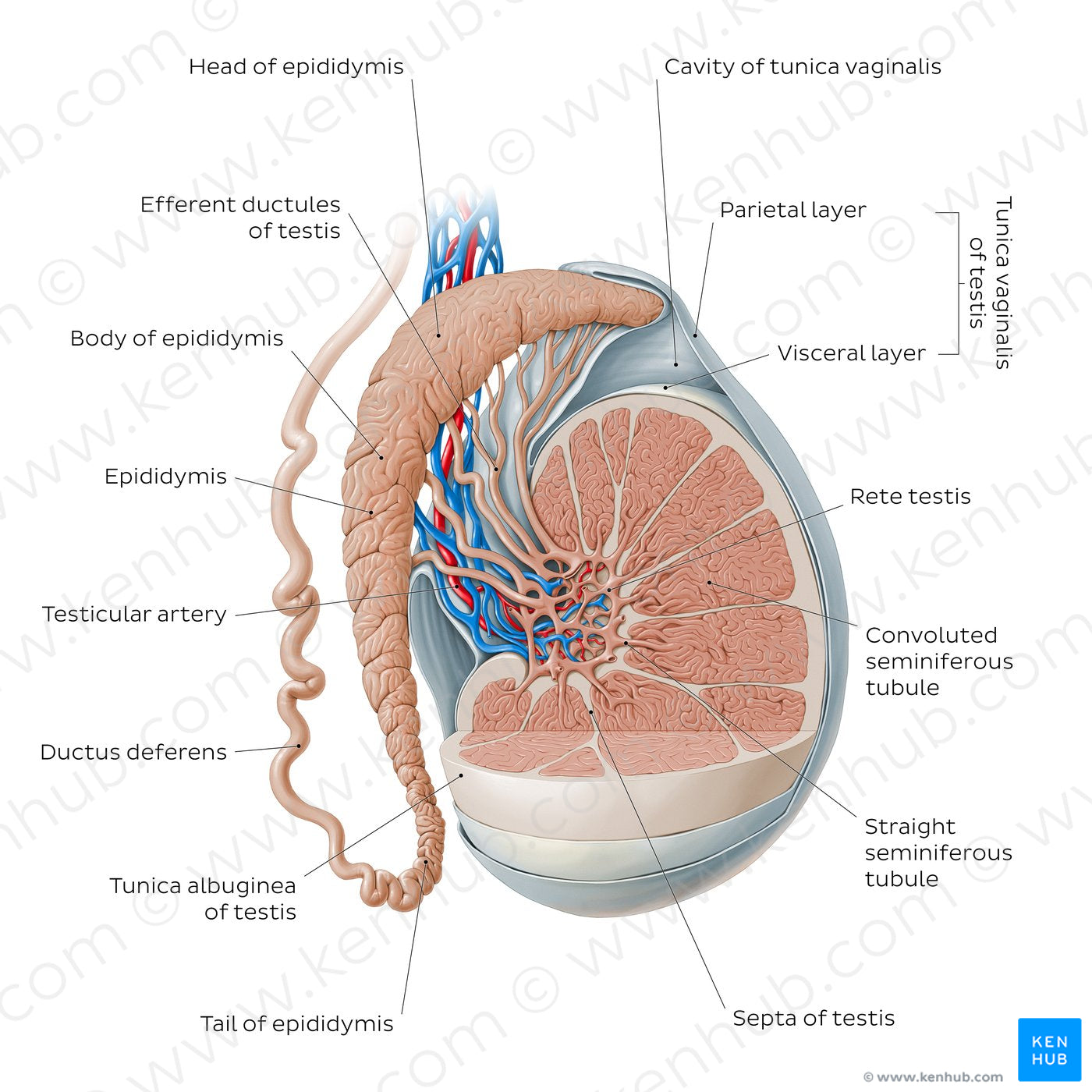
A section of the testis and surrounding connective tissue layers has been removed in order to visualize the internal structure of the testis.The convoluted seminiferous tubules form the bulk of the testis and converge to become the straight seminiferous tubules as they approach the hilum, where they form the rete testis. The efferent ductules arise from the rete testis and converge to form the head and body of the epididymis.The ductules continue to unite within the epididymis, forming a single duct, known as the tail of the epididymis, which continues as the ductus deferens. The testis is enveloped by the tunica vaginalis and the tunica albuginea. The tunica vaginalis has two layers: a visceral layer and parietal layer between which is a potential space known as the cavity of the tunica vaginalis. The tunica albuginea envelopes only the testis while the tunica vaginalis envelops the testes and much of the epididymis.
Regular price
$7.56 USD
Regular price
Sale price
$7.56 USD
Unit price
per
Couldn't load pickup availability
#0574CB
#638FAC
#1667A6
#3B3D4A
#E7B29C
#CAB7AA