
Samantha Zimmerman
Talus (Medial and lateral view) (Latin)
Talus (Medial and lateral view) (Latin)
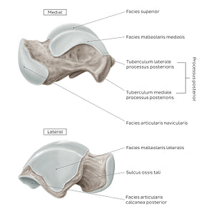
The facies articularis navicularis of the caput ossis tali is best appreciated from the medial aspect of the talus. As its name suggests, it articulates with the facies articularis ossis navicularis to form the art. talonavicularis. On the lateral view, the sulcus ossis tali of the neck (collum) is clearly seen, which forms the sinus tarsi when joined with the sulcus calcanei of the calcaneus.On the medial and lateral sides of the body, the os tali bears a facies malleolaris medialis and facies malleolaris lateralis that serve as articular surfaces for the malleoli lateralis and medialis, respectively.The facies malleolaris lateralis is a concave, triangular area that encloses the trochlea ossis tali laterally. The facies malleolaris medialis is a smooth crescent shaped area that encloses the trochlea ossis tali medially. The tuberculum mediale and tuberculum laterale of the processus posterior ossis tali provide attachment sites for the lig. talocalcaneum mediale, lig. talocalcaneum laterale and the lig. talofibulare posterius.
Regular price
$7.56 USD
Regular price
Sale price
$7.56 USD
Unit price
per
Couldn't load pickup availability

#8A7C72
#45372D
#C5BCB3