
Samantha Zimmerman
Talus (Medial and lateral view) (English)
Talus (Medial and lateral view) (English)
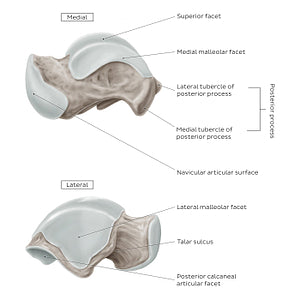
The navicular articular surface of the head of talus is best appreciated from the medial aspect of the talus. As its name suggests, it articulates with the articular surface of the navicular bone to form the talonavicular joint. On the lateral view, the talar sulcus of the neck is clearly seen, which forms the tarsal sinus when joined with the calcaneal sulcus of the calcaneus.On the medial and lateral sides of the body, the talus bears a medial and lateral malleolar facet that serve as articular surfaces for the medial and lateral malleoli, respectively. The lateral malleolar facet is a concave, triangular area that encloses the trochlea laterally. The medial malleolar facet is a smooth crescent shaped area that encloses the trochlea medially. The medial and lateral tubercles of the posterior process of the body of the talus provide attachment sites for the medial and posterior talocalcaneal ligaments and the posterior talofibular ligament.
Regular price
$7.56 USD
Regular price
Sale price
$7.56 USD
Unit price
per
Couldn't load pickup availability

#8A7C72
#45372D
#C5BCB3