
Paul Kim
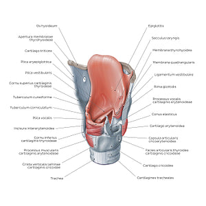
Structure of the larynx: posterolateral view (Latin)
Structure of the larynx: posterolateral view (Latin)
The epiglottis is a leaf-shaped piece of elastic cartilage attached to the facies interna of the cartilago thyroidea. When oral contents are swallowed, it folds over the aditus laryngis preventing food/fluids from entering the trachea. A thin layer of connective tissue, the membrana quadrangularis extends between the lateral borders of the epiglottis and the cartilagines arytenoideae. Its free lower edge is thickened and forms the ligamentum vestibulare. This ligament is enclosed by a fold of mucous membrane to form the plica vestibularis (false vocal cords/plicae vocales spuriae) which extends from the cartilago thyroidea to the cartilago arytenoidea. The true vocal cords (plicae vocales verae) consist of the ligamentum vocale which is the medial free edge of the conus elasticus or ligamentum thyrohyoideum laterale, as well as the musculus vocalis which comes from the medial fibers of the musculus thyroarytenoideus and the overlying mucosa which covers it.
Regular price
$7.56 USD
Regular price
Sale price
$7.56 USD
Unit price
per
Couldn't load pickup availability

#BC4B3E
#B2706D
#683029
#474448
#E29D94
#D8AFA9