
Paul Kim
Structure of the larynx: posterolateral view (English)
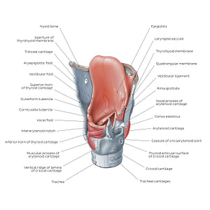
Structure of the larynx: posterolateral view (English)
The epiglottis is a leaf-shaped piece of elastic cartilage attached to the internal surface of the thyroid cartilage. When oral contents are swallowed, it folds over the laryngeal inlet preventing food/fluids from entering the trachea. A thin layer of connective tissue, the quadrangular membrane extends between the lateral borders of the epiglottis and the arytenoid cartilages. Its free lower edge is thickened and forms the vestibular ligament. This ligament is enclosed by a fold of mucous membrane to form the vestibular fold (false vocal cord) which extends from the thyroid cartilage to the arytenoid cartilage. The (true) vocal folds consist of the vocal ligament which is the medial free edge of the conus elasticus or lateral cricothyroid ligament, as well as the vocalis muscle which comes from the medial fibers of the thyroarytenoid muscle and the overlying mucosa which covers it.
Regular price
$7.56 USD
Regular price
Sale price
$7.56 USD
Unit price
per
Couldn't load pickup availability

#BC4B3E
#B2706D
#683029
#474448
#E29D94
#D8AFA9