
Paul Kim
Structure of pterygopalatine fossa (Latin)
Structure of pterygopalatine fossa (Latin)
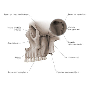
Left lateral view of the cranium with the arcus zygomaticus removed to expose the fossa pterygopalatina located between the maxilla, os sphenoidale and os palatinum. Posteriorly the fossa pterygopalatina communicates with the fossa cranii media via the foramen rotundum and canalis pterygoideus as well as with the pars nasalis pharyngis, via the canalis palatovaginalis; Medially the fissura orbitalis inferior can be seen communicating with the paries inferior cavitatis orbitalis, while laterally the fissura pterygomaxillaris connects the fossa pterygopalatina with the fossa infratemporalis. The foramen sphenopalatinum found on the medial wall of the fossa pterygopalatina opens into the lateral mucosa of the cavitas nasi.
Regular price
$7.56 USD
Regular price
Sale price
$7.56 USD
Unit price
per
Couldn't load pickup availability

#8D796C
#44382E
#C6BBB4