
Paul Kim
Structure of pterygopalatine fossa (English)
Structure of pterygopalatine fossa (English)
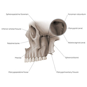
Left lateral view of the cranium with the zygomatic arch removed to expose the pterygopalatine fossa located between the maxilla, sphenoid and palatine bones. Posteriorly the pterygopalatine fossa communicates with the middle cranial fossa via the foramen rotundum and pterygoid canal as well as with the nasopharynx, via the palatovaginal canal (pharyngeal canal). Medially the inferior orbital fissure can be seen, communicating with the floor of the orbit, while laterally the pterygomaxillary fissure connects the pterygopalatine fossa with the infratemporal fossa. The sphenopalatine foramen found on the medial wall of the pterygopalatine fossa opens into the lateral mucosa of the nasal cavity.
Regular price
$7.56 USD
Regular price
Sale price
$7.56 USD
Unit price
per
Couldn't load pickup availability

#8D796C
#44382E
#C6BBB4