
Paul Kim
Spleen microcirculation (EN-LT version) (Latin)
Spleen microcirculation (EN-LT version) (Latin)
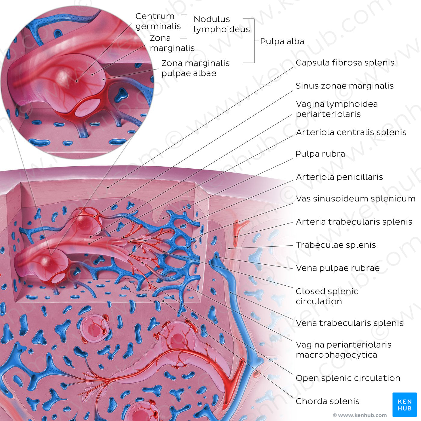
The spleen (splen) is enclosed by a fibroelastic capsule from which trabeculae extend and divide the parenchyma into sections. The majority of the parenchyma is comprised of red pulp (pulpa rubra) which is organized into splenic cords (chordae splenis) which are separated by splenic sinusoids (vasa sinusoidea splenica) where the blood is filtered from damaged red cells and cellular debris. The white pulp (pulpa alba) is composed of cylindrical formations of lymphoid tissue around a central artery (arteriola centralis); it consists of three compartments: a zona marginalis, vagina lymphoidea periarteriolaris (PALS), and noduli lymphoidei splenici. In open circulation of the spleen, blood is emptied into the chordae splenis via arteriolae penicillares where it is filtered as it passes into the vasa sinusoidea splenica. In closed circulation however, the vasa capillaria arterialia empty directly into the vasa sinusoidea splenica.
Regular price
$7.56 USD
Regular price
Sale price
$7.56 USD
Unit price
per
Couldn't load pickup availability
#3088DC
#5E6CA1
#0C3461
#583655
#EB6363
#D4A8B7