
Yousun Koh
Radius and ulna: Posterior view (English)
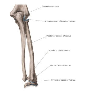
Radius and ulna: Posterior view (English)
Posteriorly, the posterior and interosseous borders of radius can be identified. The interosseous border of the radius forms the radial attachment point for the interosseous membrane of the forearm, that spans the space between the radius and ulna. The dorsal tubercle protrudes on the posterior aspect of the head of the radius and is seated between the grooves for the tendons of the extensor carpi radialis longus and brevis, as well as the tendon of the extensor pollicis longus. The styloid process of radius can also be identified from this posterior view. From a posterior aspect, the ulna is rounded and smooth and can be palpated subcutaneously along the entire length of the medial antebrachial region. The proximal end of the posterior ulna presents a hook-shaped process known as the olecranon. This bony protrusion serves as a short lever for extension of the elbow. The posterior and interosseous borders of ulna can also be appreciated from this view.
Regular price
$7.56 USD
Regular price
Sale price
$7.56 USD
Unit price
per
Couldn't load pickup availability

#8B7C73
#384752
#D1BCAE