
Yousun Koh
Posteroinferior view of the heart (Latin)
Posteroinferior view of the heart (Latin)
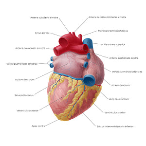
In the posteroinferior view of the heart, we can get a clear visual of the facies inferior and facies posterior (basis) cordis. The facies inferior cordis is mostly made up of the ventriculus sinister and part of the ventriculus dexter and gently slopes anteroinferiorly from the basis cordis towards the apex cordis. It is separated from the basis cordis (facies posterior) of the heart by the posterior part of the sulcus coronarius (atrioventricularis). The facies inferior is also marked by the sulcus interventricularis inferior (a.k.a. sulcus interventricularis posterior), which separates the ventriculi cordis and contains the a. interventricularis inferior (posterior) and v. cardiaca media. The facies posterior cordis is largely formed by the atrium sinistrum which is pierced by four venae pulmonales, as well a small portion of the atrium dextrum which receives the vena cava superior and inferior. The atria are separated by a shallow sulcus interatrialis, which together with the sulcus coronarius and sulcus interventricularis inferior, form the crux cordis.
Regular price
$7.56 USD
Regular price
Sale price
$7.56 USD
Unit price
per
Couldn't load pickup availability

#C98F37
#A1655D
#04355E
#6C384A
#E7CF8F
#D4ACB1