
Yousun Koh
Posteroinferior view of the heart (English)
Posteroinferior view of the heart (English)
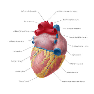
In the posteroinferior view of the heart, we can get a clear visual of the inferior and posterior surface (base) of the heart. The inferior surface is mostly made up of the left ventricle and part of the right ventricle and gently slopes anteroinferiorly from the base of the heart towards the apex. It is separated from the anatomical base (posterior surface) of the heart by the posterior part of the coronary sulcus. The inferior surface is also marked by the inferior (a.k.a. posterior) interventricular sulcus, which separates the ventricles and contains the inferior (posterior) interventricular artery and middle cardiac vein. The posterior surface is largely formed by the left atrium which is pierced by four pulmonary veins, as well a small portion of the right atrium which receives the superior and inferior venae cavae. The atria are separated by a shallow interatrial sulcus, which together with the coronary and inferior interventricular sulci, form the crux of the heart.
Regular price
$7.56 USD
Regular price
Sale price
$7.56 USD
Unit price
per
Couldn't load pickup availability

#C8374B
#A0655D
#04355E
#6F384B
#E8CF8F
#A2BFCD