
Irina Münstermann
Posterior view of the liver (Latin)
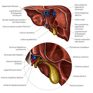
Posterior view of the liver (Latin)
The facies posterior hepatis is mainly attached to the diaphragm (diaphragma) at the area nuda hepatis; this is surrounded by the partes anterior and posterior of the lig. coronarium hepatis which merges to the left and right as the ligg. triangularia hepatis. In the center of the area nuda are vv. hepaticae dextra, media and sinistra. The lobus caudatus is limited by the pars posterior lig. coronarii, fissura lig. venosum hepatis and porta hepatis. The lobus sinister carries an impression of the stomach (impressio gastrica hepatis), while to the right of the gallbladder (vesica biliaris) is an impression of the suprarenal gland (impressio suprarenalis hepatis) and kidney (impressio renalis hepatis).
Regular price
$7.56 USD
Regular price
Sale price
$7.56 USD
Unit price
per
Couldn't load pickup availability

#1893C8
#A16D62
#024897
#54302A
#E1C86C
#D5AA9F