
Paul Kim
Overview of the oral cavity (Latin)
Overview of the oral cavity (Latin)
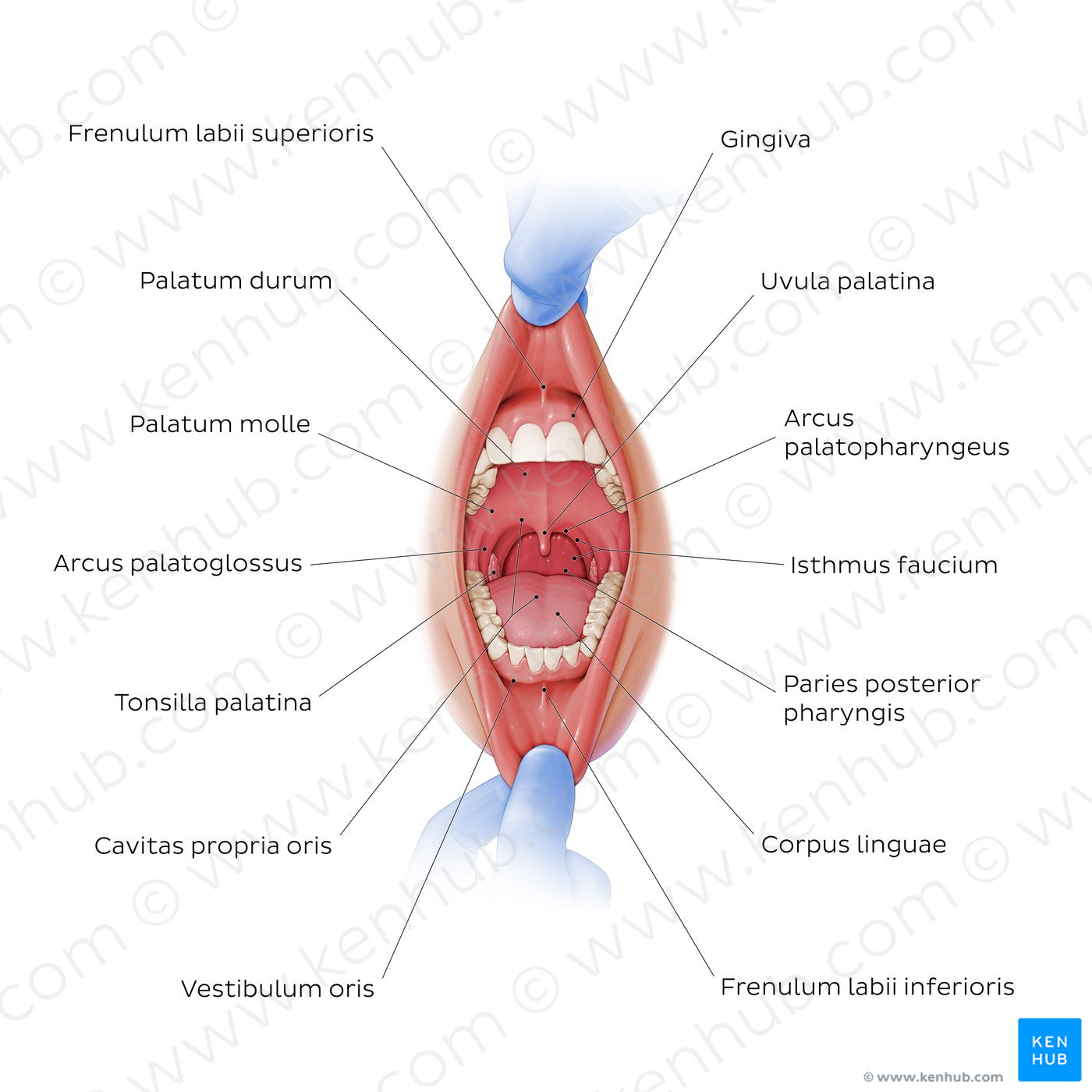
We can see the vestibulum oris with the lingual/buccal gingiva, that is firmly attached to the maxilla and mandibula, as well as the median mucosal folds known as the frenula labii superioris and inferioris. The space enclosed by the teeth is the cavitas propria oris, whose roof formed by the palatum durum and the palatum molle. The uvula palatina can be seen hanging from the posterior part of the palatum molle. The floor of that cavity is composed of the musculus geniohyoideus and musculus mylohyoideus (not shown). Sometimes the tongue (lingua) is also considered to constitute the floor of the oral cavity. Its lateral walls are formed by the dental arches (arcus dentales). Posteriorly, we can see the isthmus faucium, interposed between the cavitas oris anteriorly and the pars oralis pharyngis posteriorly. The tonsillae palatinae lie in the sinus tonsillar and is composed of lymphoid tissue, being responsible for protecting the body against organisms coming from the digestive and respiratory tracts.
Regular price
$7.56 USD
Regular price
Sale price
$7.56 USD
Unit price
per
Couldn't load pickup availability
#C43A33
#A56357
#691F22
#665355
#91B0E5
#CFADC9