
Paul Kim
Overview of the oral cavity (English)
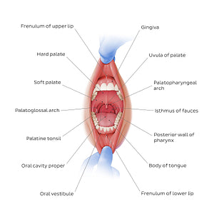
Overview of the oral cavity (English)
We can see the oral vestibule with the lingual/buccal gingiva, that are firmly attached to the maxilla and mandible, as well as the median mucosal folds known as the superior and inferior labial frenula. The space enclosed by the teeth is the oral cavity proper, whose roof is formed by the hard and soft palates. The uvula can be seen hanging from the posterior part of the soft palate. The floor of that cavity is composed of the geniohyoid and mylohyoid muscles (not shown). Sometimes the tongue is also considered to constitute the floor of the oral cavity. Its lateral walls are formed by the dental arches. Posteriorly, we can see the isthmus of fauces, interposed between the oral cavity anteriorly and the oropharynx posteriorly. The palatine tonsils lie in the tonsillar fossa (sinus) and is composed of lymphoid tissue, being responsible for protecting the body against organisms coming from the digestive and respiratory tracts.
Regular price
$7.56 USD
Regular price
Sale price
$7.56 USD
Unit price
per
Couldn't load pickup availability

#C43A33
#A56357
#662022
#5F5354
#91B0E5
#CFADC9