
Paul Kim
Optic nerve (Latin)
Optic nerve (Latin)
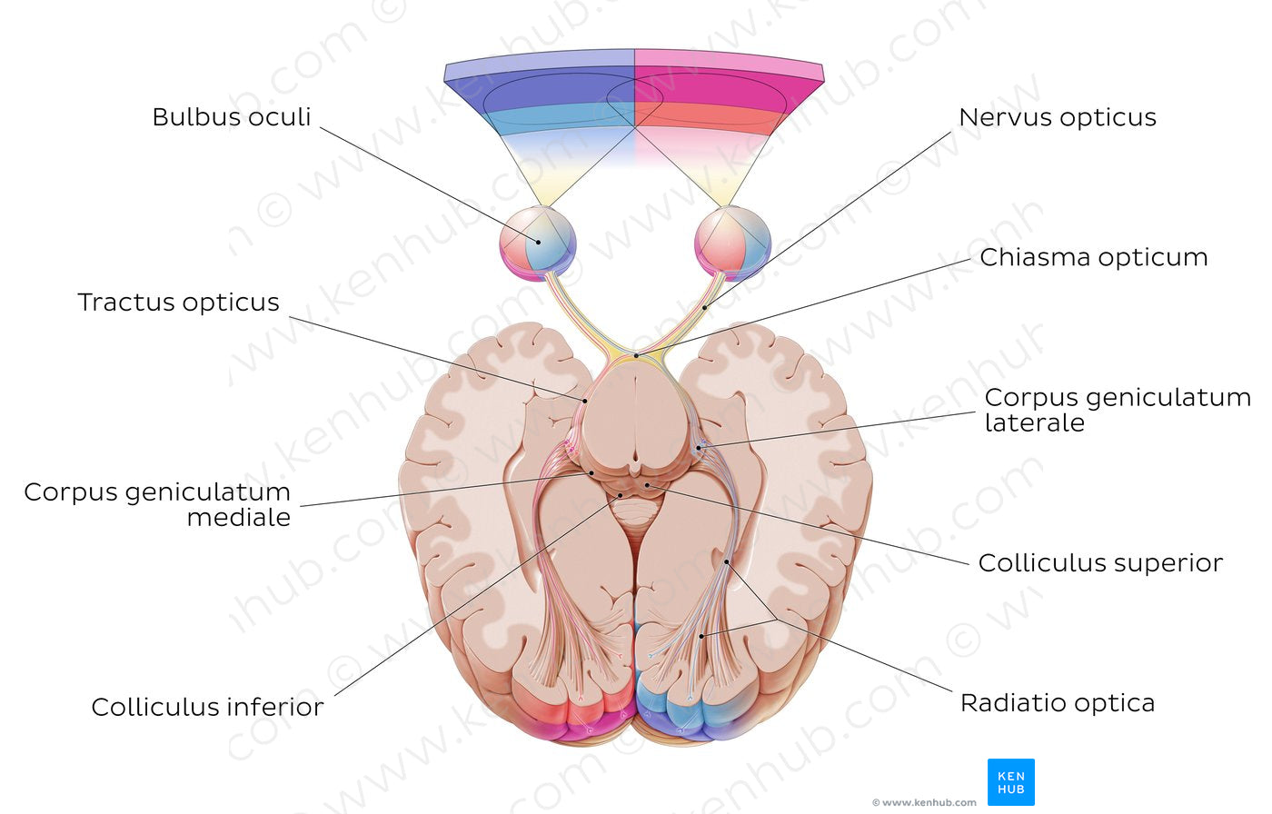
The visual pathway begins with light entering the bulbus oculi from the visual fields and being processed by the retina. Visual information is then passed on from the retina by the nervus opticus (CN II) through the canalis opticus (not shown) to the chiasma opticum in the fossa media cranii. From the chiasma opticum, the axons of the nervus opticus continue posteriorly as the tractus opticus, which then synapse at the corpus geniculatum laterale of the thalamus. Axons from the corpus geniculatum laterale travel via the radiatio optica to finally reach the primary visual cortex. It is important to note that only about 90% of the retinal axons synapse directly at the corpus geniculatum laterale. The remaining 10% project to other subcortical nuclei, mainly the colliculus superior. The colliculus superior is involved in visual reflexes, such as saccadic eye movements or tracking of objects in the visual field. It projects fibers onto the pulvinar thalami, which in turn projects onto the secondary visual cortex.
Regular price
$7.56 USD
Regular price
Sale price
$7.56 USD
Unit price
per
Couldn't load pickup availability
#E2399A
#9E5B53
#722F2B
#EC7371
#ADB3D2