
Yousun Koh
Nerves of orbit (Lateral view: eyeball in situ) (English)
Nerves of orbit (Lateral view: eyeball in situ) (English)
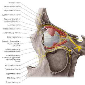
The superior and inferior branches of the oculomotor nerve (CN III) enter the orbit through the superior orbital fissure to supply the extraocular muscles of the eye. The ophthalmic nerve (CN V1) passes through the cavernous sinus and divides into three branches: the lacrimal, frontal and nasociliary nerves (not visible) before passing through the superior orbital fissure to enter the orbit. The lacrimal nerve carries sensory and autonomic nerve fibers to supply the lacrimal gland, eyelids and conjunctiva. The frontal nerve divides into supratrochlear and supraorbital branches and conveys general sensation from the forehead, glabella, frontal sinus, skin of the upper eyelid and conjunctiva. The orbit also receives partial innervation from the zygomatic and infraorbital branches of the maxillary nerve (CN V2). Located towards the posterior region of the orbit is the parasympathetic ciliary ganglion which relays parasympathetic impulses and transports sympathetic and sensory impulses to structures of the orbit.
Regular price
$7.56 USD
Regular price
Sale price
$7.56 USD
Unit price
per
Couldn't load pickup availability

#EECE21
#A35B56
#73262C
#514336
#E3D498
#C4B3AC