
Yousun Koh
Nerves of face and scalp (Lateral view) (Latin)
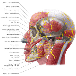
Nerves of face and scalp (Lateral view) (Latin)
The nerves providing sensory innervation to the area of the scalp posterior to the ear are the n. auricularis magnus and n. occipitalis minor (via the plexus cervicalis/rami anteriores of nervi spinales C2/C3), as well as the nn. occipitales major et tertius (which arise from the posterior ramus of spinal nerve C2). The area of the scalp anterior to the ears is innervated by the n. zygomaticotemporalis, a branch of the n. maxillaris (CN V2) and the n. auriculotemporalis, which is a branch of the n. mandibularis (CN V3). As discussed in the last image, the anterior scalp is innervated by the n. supratrochlearis and n. supraorbitalis, which are branches of the n. ophthalmicus (CN V1).
Regular price
$7.56 USD
Regular price
Sale price
$7.56 USD
Unit price
per
Couldn't load pickup availability

#EDD223
#A9625C
#78272D
#5C2C2E
#E4D198
#CBB3B0