Yousun Koh
Muscles of the pharynx (Latin)
Muscles of the pharynx (Latin)
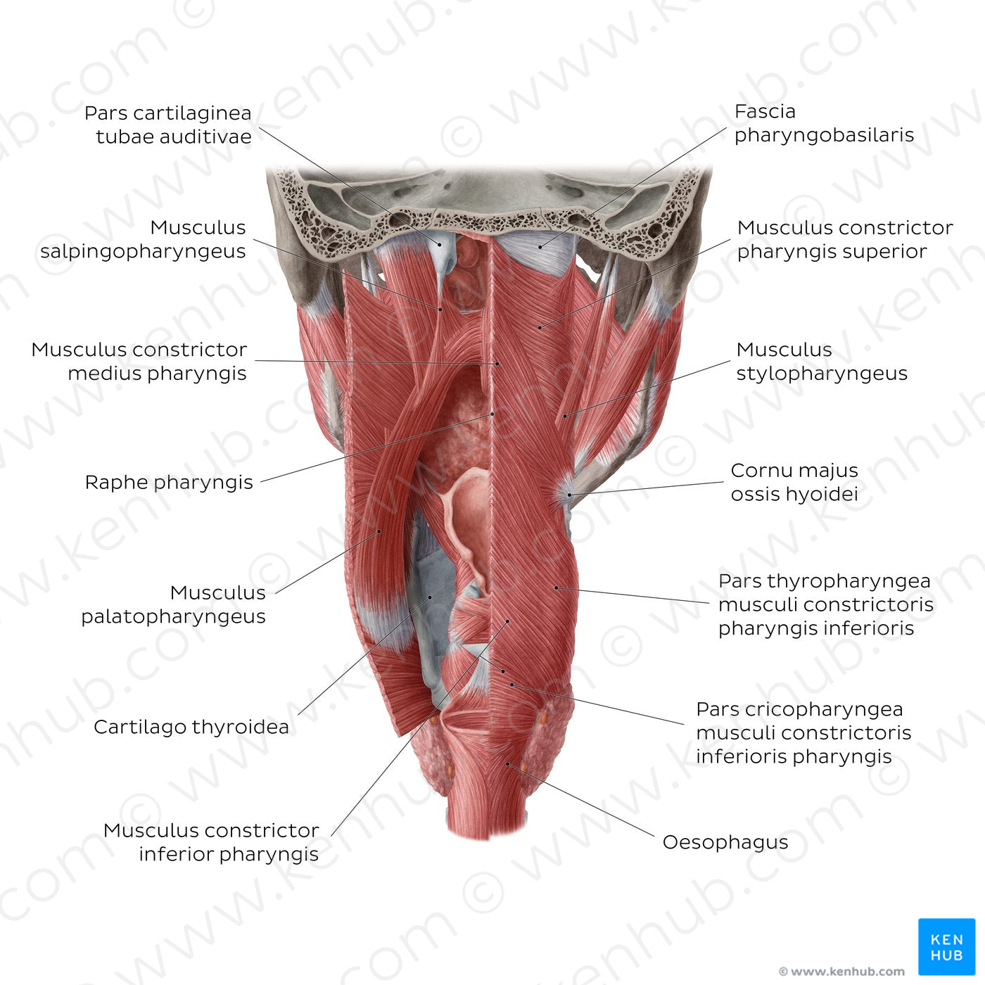
The mm. constrictores pharyngis on either side of the pharynx are the main components of the pharyngeal wall. These muscles are named according to their position: m. constrictor superior, medius et inferior. The m. constrictor inferior is further divided into a pars thyropharyngea and pars cricopharyngea. Posteriorly, these muscles come together in the midline at the raphe pharyngis. The muscular sleeve formed by these muscles has a strong internal lining known as the fascia pharyngobasilaris which is particularly evident superior to the level of the m. constrictor superior - here the pharyngeal wall is formed almost completely of fascia. The mm. elevatores/longitudinales pharyngis are located deep to their circular counterparts and include the m. stylopharyngeus, the m. salpingopharyngeus and the m. palatopharyngeus which originate from the processus styloideus ossis temporalis, tuba auditiva, and palatum molle, respectively.
Regular price
$7.56 USD
Regular price
Sale price
$7.56 USD
Unit price
per
Couldn't load pickup availability


#B0504E
#AA5754
#601915
#493831
#E2A6A2
#CDB3AE

