
Yousun Koh
Muscles of the pelvic floor (Latin)
Muscles of the pelvic floor (Latin)
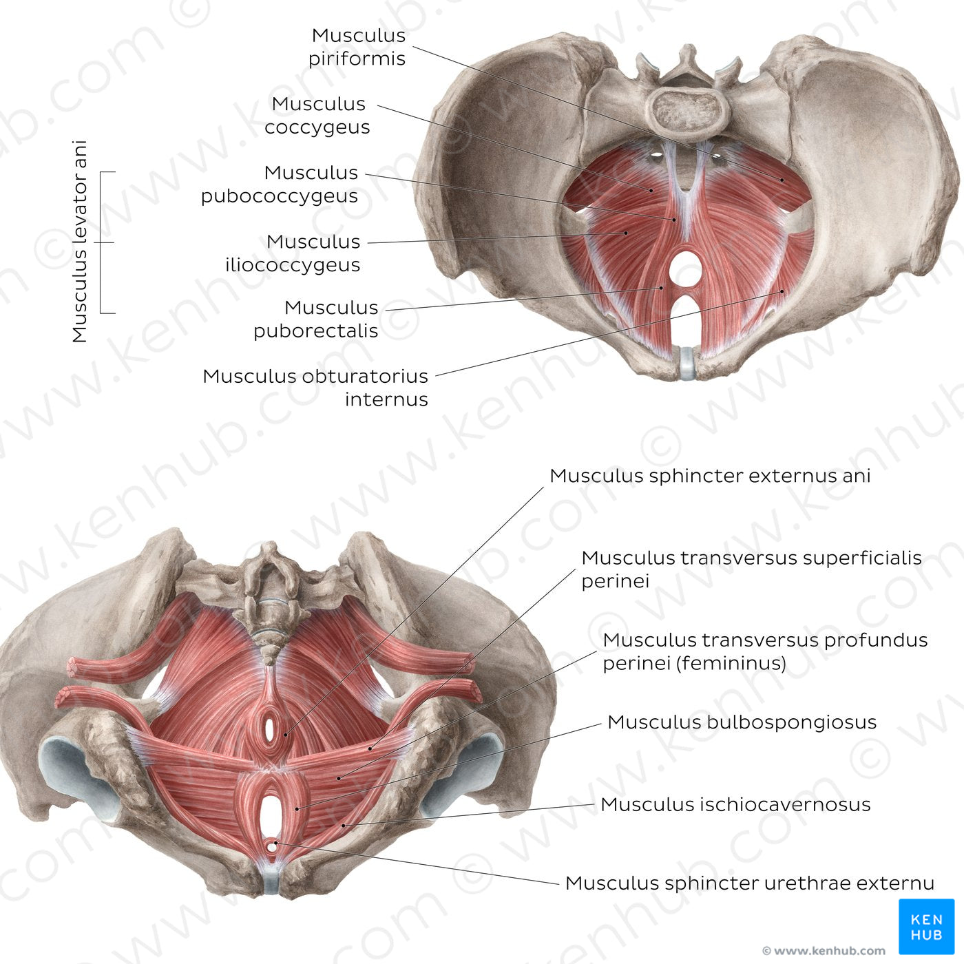
The upper image shows the superior view of the pelvic floor, while the lower image demonstrates the inferior view. From the superior view, the m. levator ani and its three components are visible: which is composed of the m. puborectalis (m. puboanalis), m. pubococcygeus, and m. iliococcygeus. Extending between the ischial spines and coccyx is the m. coccygeus. Moving posterosuperiorly, the m. piriformis forms the posterolateral wall of the pelvic cavity, while the m. obturatorius internus forms part of the anterolateral wall of the pelvic cavity. From the inferior view, the mm. perinei can be identified. Beginning in the spatium profundum perinei, there is the m. transversus profundus perinei and the m. sphincter externus urethrae. Moving inferiorly to the spatium superficiale perinei, the m. transversus superficialis perinei, m. bulbospongiosus, and paired m. ischiocavernosus are found. Finally, heading posteriorly to the anal triangle (trigonum anale) of the perineum, the m. sphincter externus ani is depicted.
Regular price
$7.56 USD
Regular price
Sale price
$7.56 USD
Unit price
per
Couldn't load pickup availability
#C6665D
#A75953
#571D19
#4F3731
#E69F9A
#CFAFAC