Yousun Koh
Muscles of the anterior neck (English)
Muscles of the anterior neck (English)
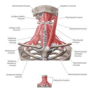
The upper image shows the platysma. This sheet-like muscle lies most superficially within the subcutaneous tissue and covers all of the anterior aspect of the neck. The lower image shows all other anterior neck muscles that are situated deep to the platysma. The sternocleidomastoid is a two-headed muscle and can be prominently seen and palpated along the lateral sides of the neck creating a ‘V-shape’. The suprahyoid (digastric, mylohyoid, geniohyoid and stylohyoid) and infrahyoid (sternohyoid, omohyoid, sternothyroid and thyrohyoid muscle) muscles position the hyoid bone, thus playing an active role in swallowing and the movement of the larynx. The scalene muscles (scalenus anterior, middle and posterior) attach to the upper two ribs, making them accessory muscles of respiration. The prevertebral muscles (rectus capitis anterior, rectus capitis lateralis, longus capitis and longus colli) are located along the length of the anterior cervical spine and are surrounded by the prevertebral fascia of the neck. These muscles help with the flexion of the head to varying degrees.
Regular price
$7.56 USD
Regular price
Sale price
$7.56 USD
Unit price
per
Couldn't load pickup availability


#AE4B4B
#9E5655
#8A1A1B
#4A3A33
#E49393
#C8B2AC

