
Paul Kim
Motor and sensory cortical homunculus (English)
Motor and sensory cortical homunculus (English)
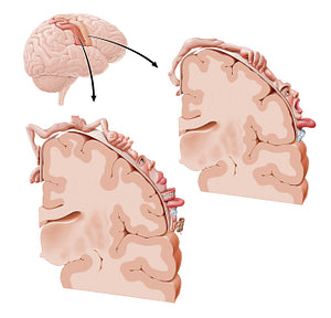
This image shows coronal sections of the brain through the precentral and postcentral gyri. The motor homunculus (upper right image) represents the topographic map of the motor innervation of the body. Note that the body parts of the homunculus are not proportional to the real body parts. This is because the amount of cortex dedicated to each body part is proportional to the intricacy and complexity of the motor function of each body part. The sensory homunculus (lower left image) is a topographic distribution of the somatosensory innervation of different body parts. Again, the area of the cortex that is responsible for the innervation of the body parts, is not proportional to the dimensions of the body part. The amount of cortex per body part is proportional to the complexity of sensations received from that organ.
Regular price
$7.56 USD
Regular price
Sale price
$7.56 USD
Unit price
per
Couldn't load pickup availability

#C35D3A
#B26A53
#691C0E
#6E4635
#E4A78E
#D3B6AE