
Mao Miyamoto
Middle ear: Sagittal section (English)
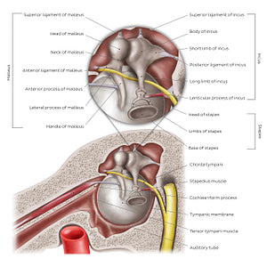
Middle ear: Sagittal section (English)
This sagittal section (medial view) of the middle ear provides a better view of the three auditory ossicles, their parts and their ligaments. The malleus consists of a head, neck, anterior and lateral processes and handle. It is suspended via three ligaments: the superior, anterior and lateral ligaments of malleus (the former of which is not seen on this view). The incus consists of a body, short and long limbs, and lenticular process. It is suspended by two ligaments: the superior and posterior ligaments of incus. The stapes consists of a head, anterior and posterior limbs, and base; it is suspended via the anular ligament of stapes (not shown). In addition, the tensor tympani muscle is clearly seen on this view, running through the semicanal for the tensor tympani muscle of the temporal bone across the cochleariform process which acts as a pulley for this muscle. This view also provides a visual of the stapedius muscle, which attaches on the neck of the stapes.
Regular price
$7.56 USD
Regular price
Sale price
$7.56 USD
Unit price
per
Couldn't load pickup availability

#CD2F2D
#A5645B
#5B2118
#53342D
#EEE59B
#D2CEA8