Paul Kim
Meninges of the brain (coronal section) (Latin)
Meninges of the brain (coronal section) (Latin)
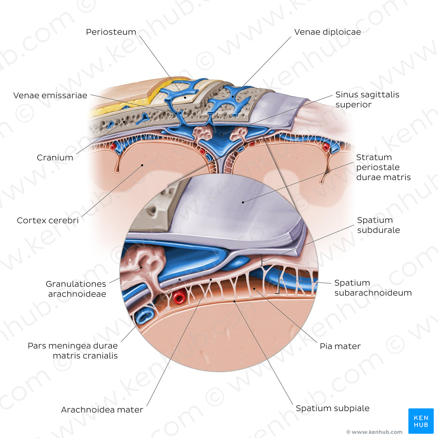
Coronal section through a portion of the skull, meninges and cerebral cortex. The cranial dura mater is composed of two layers: the outer stratum periosteale (which adheres tightly to the cranium, also known as the endocranium) and inner stratum meningeale. The two dural layers are usually directly apposed to each other, except in places where they separate to form the sinus venosi durales, which is represented in this image by the sinus sagittalis superior. Deeper to the dura mater, is the arachnoid mater with its small protrusions known as granulationes arachnoideae, that pierce the inner layer of the dura projecting into the lumen of the sinus sagittalis superior. The pia mater is a thin membrane composed of a single cell layer which, unlike the dura and arachnoid mater, closely follows all contours (i.e. gyri and sulci) of the brain. Thin projections of connective tissue called trabeculae arachnoideae extend from the inner surface of the arachnoid mater, traverse the spatium subarachnoidale and attach to the outer surface of the pia mater. The spatium subdurale is a potential space that can be opened by the separation of the arachnoid from the dura as a result of trauma or other ongoing pathological process; in an healthy state both meninges are attached to each other and the spatium subdurale is obliterated. The spatium subarachnoidale, between the arachnoid and pia mater, is a fluid-filled space occupied by CSF. Cerebral blood vessels pass through this space, however are enveloped in a layer of leptomeningeal epithelial cells (not shown in image) and thus are not in contact with the CSF. Underneath the pia mater is the thin spatium subpiale which separates it from the glia limitans of the cerebral cortex.
Regular price
$7.56 USD
Regular price
Sale price
$7.56 USD
Unit price
per
Couldn't load pickup availability


#1F78CD
#A2675B
#16528F
#6F4538
#7EC5FB
#D1B5AB

