
Esther Gollan
Lymphatics of the posterior abdominal wall (Latin)
Lymphatics of the posterior abdominal wall (Latin)
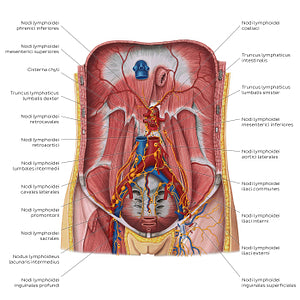
Illustration of the paries posterior abdominis and cavitas pelvica, with the organs and other structures removed to reveal the lymph nodes and neighbouring vessels. In the center of the figure, adjacent to the aorta abdominalis, is the cisterna chyli, the main drainage pathway of the paries posterior abdominis. Two trunks converge to form the cisterna chyli: the truncus lymphaticus lumbalis dexter and truncus lymphaticus lumbalis sinister (which usually receives the truncus lymphaticus intestinalis). These structures receive the lymph mainly from the nll. lumbales (which are composed of the nll. lumbales sinistri, also known as nll. aortici and the nll. lumbales dextri, also called nll. cavales) as well as the nll. celiaci et mesenterici superiores/inferiores which drain the canalis gastrointestinalis. Pelvic organs drain to the nll. Iliaci communes/interni/externi, which in turn drain to nll. lumbales.
Regular price
$7.56 USD
Regular price
Sale price
$7.56 USD
Unit price
per
Couldn't load pickup availability

#DE952F
#A95756
#641819
#562A2A
#E6C793
#D2AFAD