Yousun Koh
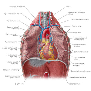
Lungs in situ (English)
Lungs in situ (English)
In this image, both the right and the left lungs are slightly retracted so the relations of the lungs with the heart and the mediastinal structures can be seen. The right lung lies in close relation to the superior and inferior venae cavae as well as the azygos vein (not seen), while the left lung lies in close approximation with all parts of the thoracic aorta. Both lungs conform to the shape of the heart. Each lung is suspended from the mediastinum by its root: a pedicle formed by structures entering and exiting the lungs via the hilum (e.g., bronchi, pulmonary/bronchial vasculature, lymphatics and nerves).Notice the two potential spaces (out of four in total) between the lungs and the parietal pleura, also known as the pleural recesses. The costodiaphragmatic recess is located at the inferior most part of the pleural cavity whereas the costomediastinal recess lies anteriorly, between the costal and mediastinal layers of parietal pleura. Those recesses are usually empty, so dull percussion sound in those areas as well as positive chest radiograph can indicate a pathological condition.
Regular price
$7.56 USD
Regular price
Sale price
$7.56 USD
Unit price
per
Couldn't load pickup availability


#CC3D3D
#AB5452
#6A201E
#5B395D
#E49898
#D3AEAB

