
Irina Münstermann
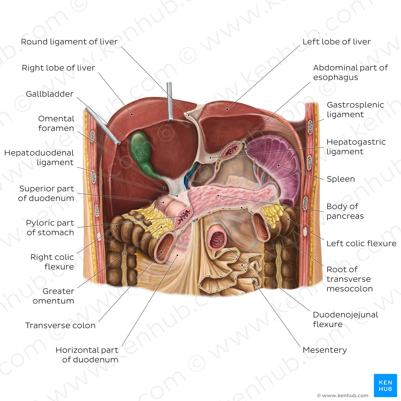
Liver in situ (English)
Liver in situ (English)
Anterior view of the abdomen with the liver retracted and the stomach removed to expose the underlying structures. Right and left lobes of the liver can be seen with the gallbladder on the posterior surface of the right lobe and with the blood vessels and the bile duct enclosed in the hepatoduodenal ligament (part of the lesser omentum). The rest of the lesser omentum is formed by the hepatogastric ligament. It extends between the liver and the stomach, but only a small part of it is visible around the cardiac orifice of the esophagus. The falciform ligament is located between the right and left lobes and is continuous inferiorly with the round ligament of the liver, which is exposed due to the reflected liver. The round ligament extends posteroinferiorly to join the ligamentum venosum. A section of the greater omentum, the superior part of the duodenum and the transverse colon are all shown and form the posteroinferior anatomical relations of the liver. Not shown in the image are the posterior relations of the liver to the right kidney and the suprarenal gland.
Regular price
$7.56 USD
Regular price
Sale price
$7.56 USD
Unit price
per
Couldn't load pickup availability
#C39038
#AE6F53
#67281C
#5A4931
#E9B390
#D4B3AB