
Paul Kim
Lens and corpus ciliare: Posterior view (English)
Lens and corpus ciliare: Posterior view (English)
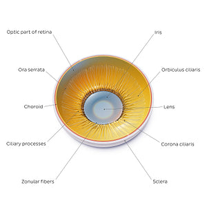
At the centre of this image is the lens, located posterior to the iris and anterior to the postremal/vitreous chamber of the eye. The capsule of the lens is anchored to adjacent ciliary processes of the ciliary body by zonular fibres which collectively form the suspensory ligament of the lens/ciliary zonule. The anterior portion of the ciliary body is known as the corona ciliaris/pars plicata and is marked by ciliary processes (separated by ciliary folds) which function to produce aqueous humour within the posterior chamber providing nutrients for the cornea and lens. The orbiculus ciliaris/pars plana forms the posterior portion of the ciliary body and terminates along the ora serrata. The optic part of the retina is continuous with the choroid and sclera before terminating anteriorly at the ora serrata while the non-visual part of the retina extends over the ciliary body and iris.
Regular price
$7.56 USD
Regular price
Sale price
$7.56 USD
Unit price
per
Couldn't load pickup availability

#E6A918
#A29059
#5D330E
#8E764E
#EAD58A
#D6A5A1