
Paul Kim
Knee joint - sagittal (Latin)
Knee joint - sagittal (Latin)
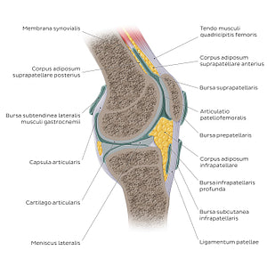
Sagittal view of the right knee joint (art.genus), with all articulating surfaces clearly visible. The condylus lateralis and condylus medialis ossis femoris articulate with the planum tibiae inferiorly forming the tibiofemoral joint (art. tibiofemoralis). Anteriorly, the facies patellaris ossis femoris articulates with the facies articularis patellae forming the patellofemoral joint (art. patellofemoralis). This view of the articulatio genus is best for examining the structure of the capsula articularis and its two parts, the outer stratum fibrosum, and inner stratum synoviale which encloses the cavitas articularis. The capsula articularis forms several pouches called bursae, that cushion and reduce friction within the art. genus. Additional important structures are the menisci situated between the condylus medialis and lateralis ossis femoris and planum tibiae, providing congruency to these articulating surfaces. In the art. patellofemoralis, one of the important supporting structures is the ligamentum patellae, that extends from the patella to the tuberositas tibiae.
Regular price
$7.56 USD
Regular price
Sale price
$7.56 USD
Unit price
per
Couldn't load pickup availability

#E0AB37
#6D9499
#E0AB37
#385B57
#EED693
#ACBAD5