Yousun Koh
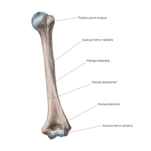
Humerus: Posterior view (Latin)
Humerus: Posterior view (Latin)
Due to its lateral positioning, the tuberculum majus humeri can also be identified from this posterior view. Posteriorly, the shaft of the humerus is marked by the oblique sulcus nervi radialis, which allows for the passage of the n. radialis and a. profunda brachii. In addition, the margo lateralis and facies posterior of the humerus can be appreciated from the posterior view. The distal end of the posterior humerus presents with a large fossa known as the fossa olecrani. In elbow extension, the tip of the ulnar olecranon process lodges into this fossa. The epicondylus medialis of the humerus contains a shallow ridge on its posterior surface, known as the sulcus nervi ulnaris. As its name suggests, the groove transmits the n. ulnaris.
Regular price
$7.56 USD
Regular price
Sale price
$7.56 USD
Unit price
per
Couldn't load pickup availability


#8D796F
#354956
#E0CABD
#D1BAAD

