Yousun Koh
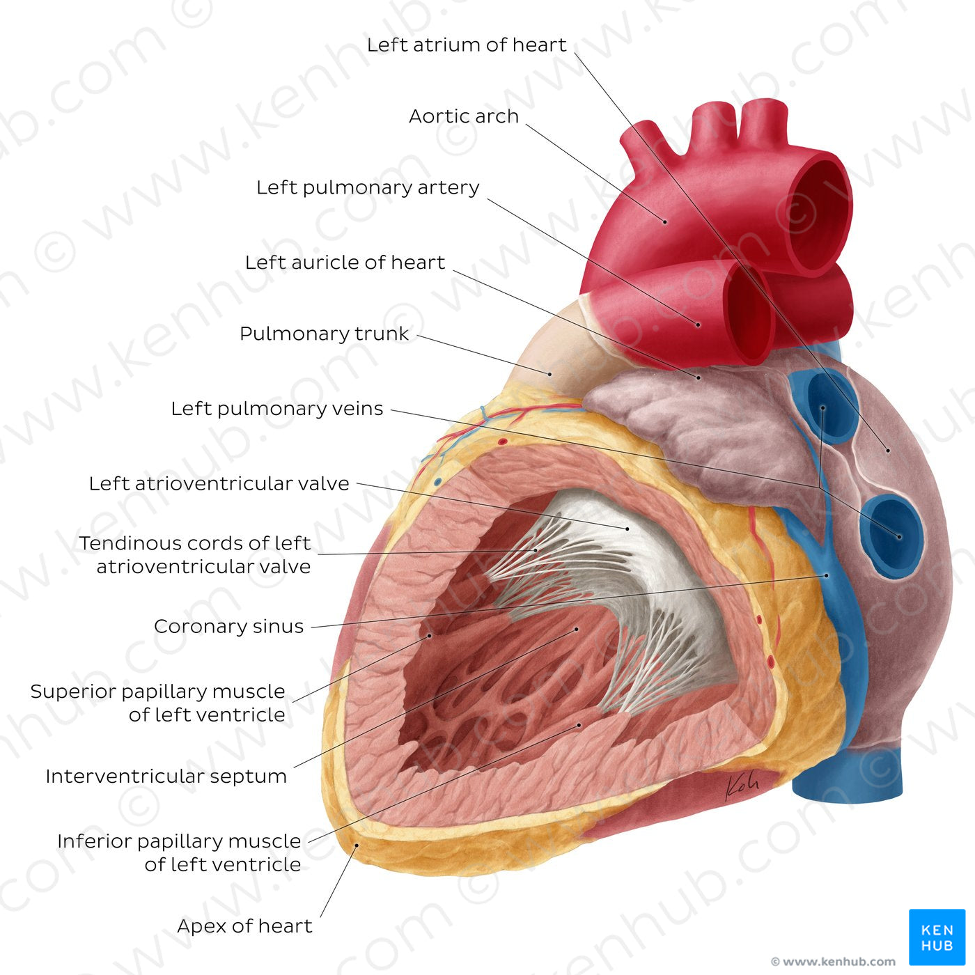
Heart: Left ventricle (English)
Heart: Left ventricle (English)
The left ventricle has noticeably thicker muscular walls which facilitate the generation of sufficient force to overcome the higher blood systemic pressure of the aorta. Similar to its right counterpart, the internal structure of the ventricle features notable muscular ridges, known as trabeculae carneae. The left atrioventricular (a.k.a. mitral) valve has two leaflets, anterior and posterior. Each of these are connected to a corresponding superior (or anterior) and inferior (or posterior) papillary muscle via tendinous cords, also known as chordae tendineae. A small conical projection of the left atrium, known as the left auricle (or atrial appendage) can be seen adjacent to the root of the pulmonary trunk. The posterior wall of the left atrium is also pierced by four pulmonary veins which carry oxygenated blood from the lungs.
Regular price
$7.56 USD
Regular price
Sale price
$7.56 USD
Unit price
per
Couldn't load pickup availability


#C7364B
#5388AB
#6F0508
#74443E
#F0CF91
#CDB1AD

