
Liene Znotina
Femur (posterior view) (Portuguese)
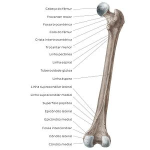
Femur (posterior view) (Portuguese)
O trocânter menor é facilmente observado na vista posterior, separado do trocânter maior pela crista intertrocantérica. A crista intertrocantérica e a linha intertrocantérica (na vista anterior) marcam a transição entre o colo e a diáfise do fêmur. Na superfície medial do trocânter maior encontra-se uma depressão em forma de crescente, conhecida como fossa trocantérica. Inferiormente ao trocânter menor, encontram-se três cristas ósseas, chamadas de linha pectínea, linha espiral e tuberosidade glútea. Essas três cristas convergem inferiormente para formarem a linha áspera, que segue ao longo de toda a extensão da diáfise femoral, até se dividir em duas cristas em sua extremidade distal: as linhas supracondilares medial e lateral. A superfície posterior da extremidade distal do fêmur permite visualizar melhor os côndilos medial e lateral, os quais, nesta vista, se encontram separados pela fossa intercondilar.
Regular price
$7.56 USD
Regular price
Sale price
$7.56 USD
Unit price
per
Couldn't load pickup availability

#8F806D
#57473C
#C7BCB3