
Liene Znotina
Femur (posterior view) (Latin)
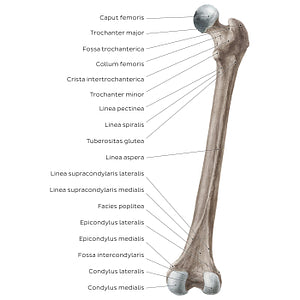
Femur (posterior view) (Latin)
The trochanter minor is clearly seen on the posterior view, separated from the trochanter major by the crista intertrochanterica. The crista intertrochanterica and linea intertrochanterica (on the anterior view) mark the transition between the collum and corpus ossis femoris. Found on the medial surface of the trochanter major is the crescent-shaped depression known as the fossa trochanterica. Seen below the trochanter minor are the 3 small bony ridges called linea pectinea, linea spiralis and tuberositas glutea. These 3 ridges converge inferiorly to form the linea aspera that runs along the entire corpus ossis femoris. Near the distal end of the femur, the linea aspera diverges into 2 ridges: the linea supracondylaris medialis and lateralis. The posterior surface of the distal end of the femur provides a better visual of the condylus medialis and lateralis, which in this view are separated by the fossa intercondylaris.
Regular price
$7.56 USD
Regular price
Sale price
$7.56 USD
Unit price
per
Couldn't load pickup availability

#8F806D
#57473C
#C7BCB3