Liene Znotina
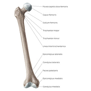
Femur (anterior view) (Latin)
Femur (anterior view) (Latin)
The anterior view of the femur features several important landmarks. The most proximal portion of the caput ossis femoris features a small dimple, known as the fovea capitis ossis femoris. Below the collum ossis femoris are the trochanter major and minor, with the linea intertrochanterica spanning between them. These bony prominences act as important attachment sites for the muscles of the hip and thigh. The distal end of the femur contains the condylus medialis and lateralis that articulate with the tibia, as well as the epicondylus medialis and lateralis above them. Between the condylus medialis and lateralis is the facies patellaris ossis femoris, which as its name suggests, articulates with the patella, contributing to the formation of the articulatio genu (knee joint). Notice how some of these landmarks are better seen on the posterior view (2nd image), such as the trochanter minor, and the condylus medialis and lateralis.
Regular price
$7.56 USD
Regular price
Sale price
$7.56 USD
Unit price
per
Couldn't load pickup availability


#8C7D72
#483A32
#C8BCB3

