
Irina Münstermann
Female pelvic viscera and perineum (English)
Female pelvic viscera and perineum (English)
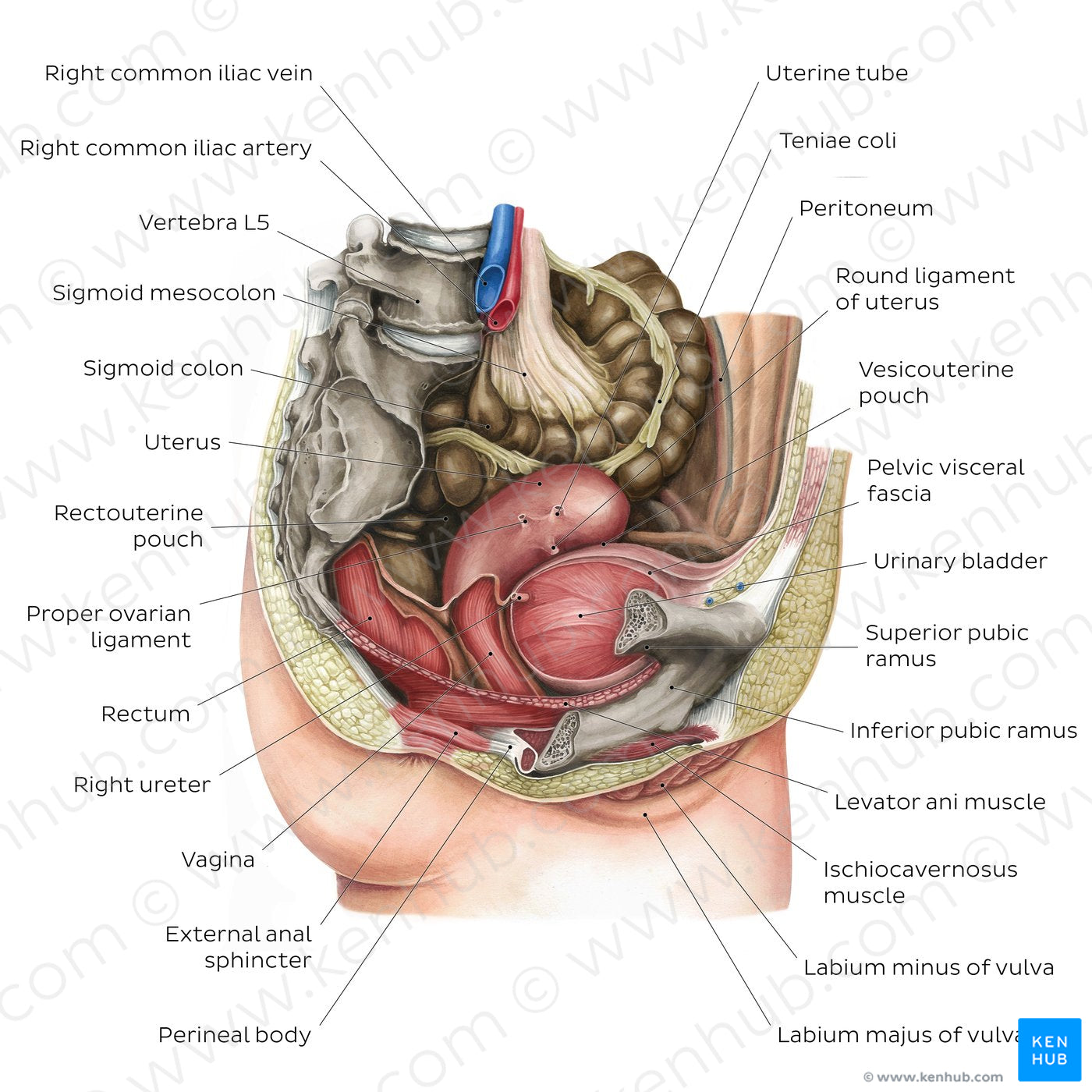
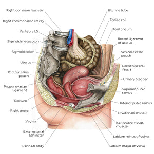
Parasagittal section of the female pelvis and perineum (right view) with parts of the right pelvic wall, fascia, ovary and other structures removed to show the relations of the female pelvic organs. The urinary bladder is located anteriorly, just behind the pubic bone, with the distal ‘stump’ of the right pelvic ureter at its base. The uterus sits between the urinary bladder and the rectum and shows stumps of the right proper ovarian ligament, uterine tube, and round ligament of the uterus. The vagina extends from the inferior cervical region of the uterus and opens into the perineum at the vaginal orifice (opening). The terminal part of the sigmoid colon continues distally as the rectum, both of which are located in the posterior aspect of the pelvic cavity, anterior to the sacrum and coccyx.
Regular price
$7.56 USD
Regular price
Sale price
$7.56 USD
Unit price
per
Couldn't load pickup availability
#A22831
#9E8B59
#54271C
#4C372A
#E8B095
#D3CAB0