
Paul Kim
Eyeball (English)
Eyeball (English)
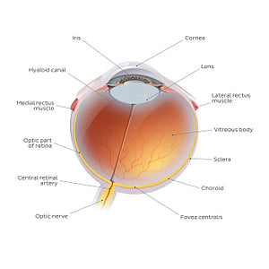
The choroid of the eye is located between the sclera and retina and is filled with numerous vascular bundles which supply the outer portion of the retina. Posterior to the iris and anterior portion of the sclera is a thickening of muscular and connective tissue known as the ciliary body. The final component of the vascular layer is the pigmented iris which contains a central aperture known as the pupil. The retina forms the inner layer of the eye and is composed of non-visual and optic parts. Located posterior to the lens and enveloped by the retina is a compartment known as the postremal/vitreous chamber which is occupied by a semi-solid/jelly-like structure known as the vitreous body. Embedded within the meshes of the vitreous body is a fluid-like substance known as vitreous humor. Together these structures allow for the passage of light to the retina and provide structural support to the lens anteriorly.
Regular price
$7.56 USD
Regular price
Sale price
$7.56 USD
Unit price
per
Couldn't load pickup availability

#EBCA3B
#966861
#A03D22
#523B33
#F2CD72
#D1B2B1