Paul Kim
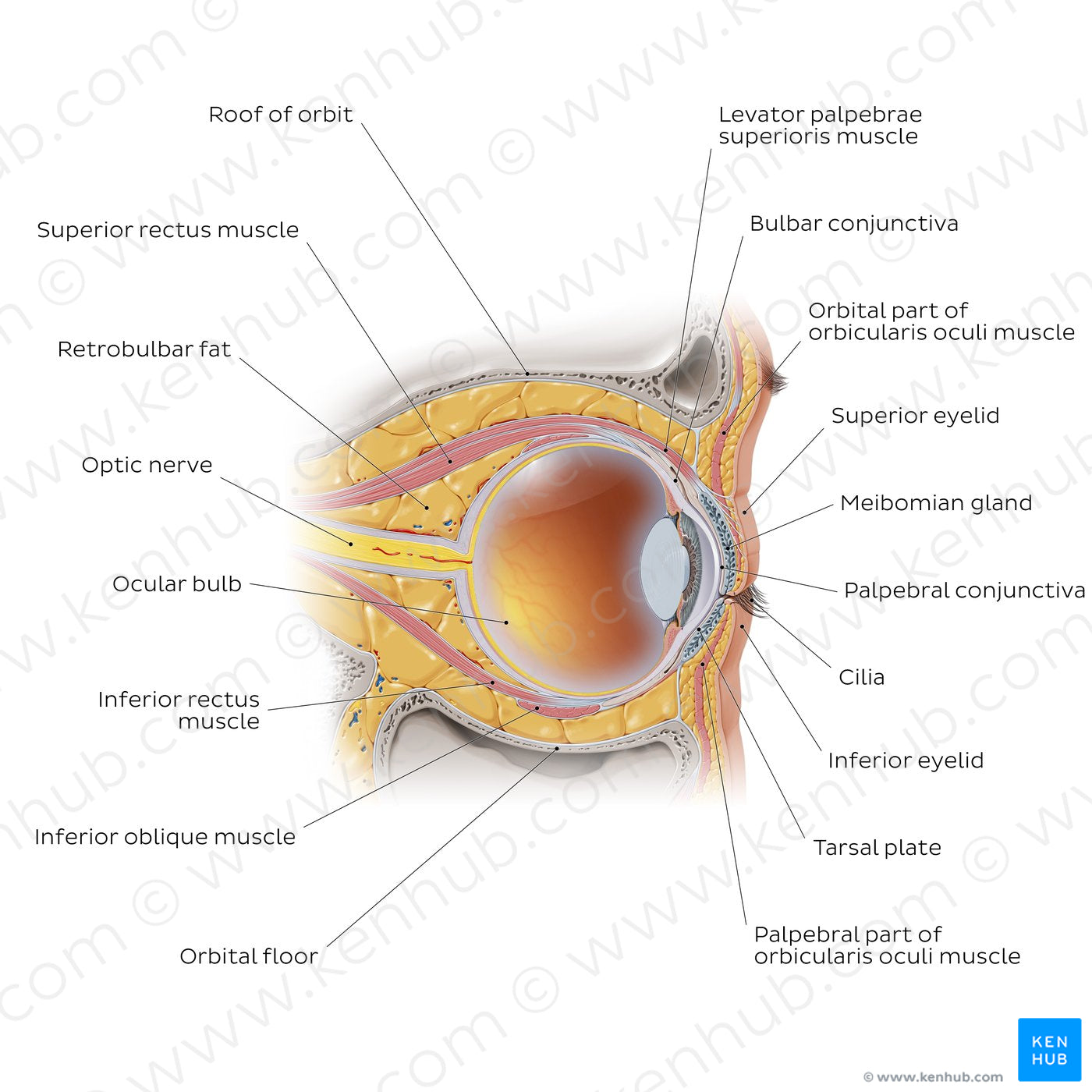
Eye in situ: sagittal section (English)
Eye in situ: sagittal section (English)
This image allows an appreciation of a number of accessory visual structures which support and protect the eyeball. The eyeball is suspended within the anterior portion of the orbit by six extraocular muscles and by a fascial sheath. There are seven extraocular muscles altogether which can be divided into four recti, two obliques and one levator palpebrae superioris muscle. The levator palpebrae superioris muscle attaches to the upper eyelid and functions to raise and maintain it in an open position. The other six extraocular muscles extend to insert onto the sclera of the eye and are responsible for controlling movement. Lying against the anterior aspect of the eyeball are the palpebral conjunctiva and eyelids (as well as their associated glands) which protect the eyeball from the external environment. Surrounding the eyeball is a mass of adipose tissue known as retrobulbar fat. It functions to cushion and support the eyeball within the orbit and prevent excessive posterior pull on the eyeball by the rectus muscles. Extending from the posterior aspect of the eyeball is the central retinal artery and vein which pierces the optic nerve before it reaches the eyeball. The optic nerve exits the orbit through the optic canal and transmits sensory information from the eye to the brain.
Regular price
$7.56 USD
Regular price
Sale price
$7.56 USD
Unit price
per
Couldn't load pickup availability


#C5370F
#A66957
#8A3C20
#4F3D36
#EACD8E
#CAB6B0

