
Begoña Rodriguez
Duodenum (Latin)
Duodenum (Latin)
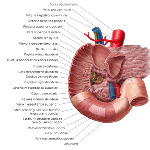
The anterior wall of the pars superior duodeni and pars descendens duodeni is removed to reveal its internal structure. Plicae circulares of mucosa cover most of its luminal surface and two small openings are seen amongst them in the pars descendens duodeni. The papilla major duodeni allows pancreatic enzymes and bile to enter the duodenum from the union of the ductus pancreaticus and ductus biliaris, known as the ampulla hepatopancreatica. The papilla minor duodeni is an opening for pancreatic enzymes from the ductus pancreaticus accessorius. The different layers of the duodenum are exposed in the pars horizontalis duodeni. The caput pancreatis sits within the curve of the duodenum. The aorta abdominalis, vena porta hepatis (pictured), v. cava inferior, arteriae pancreaticoduodenales, ductus biliaris, right kidney (ren dexter), ureter dexter, musculus psoas major dexter, a. and v. ovarica/testicularis and vertebra L3 (not pictured) are located posterior to the duodenum. The a. and v. mesenterica superior (pictured), lobus dexter hepatis and vesica biliaris extend over the anterior surface of the duodenum.
Regular price
$7.56 USD
Regular price
Sale price
$7.56 USD
Unit price
per
Couldn't load pickup availability

#DF1926
#A85957
#631C1B
#5A3D2B
#E89591
#D3ACA9