Irina Münstermann
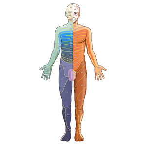
Dermatomes: Foerster/Keegan and Garrett map - Anterior (English)
Dermatomes: Foerster/Keegan and Garrett map - Anterior (English)
The Foerster (right) and Keegan and Garrett (K&G; left) dermatome maps of the ventral surface of the body side by side. There are three dermatomes of the face, V1, V2 and V3, which are innervated by the three branches of the trigeminal nerve (CN V), and have a similar distribution in both dermatome maps. A slight difference between the two maps is seen in the neck and shoulder regions, which on the Foerster map, are represented as C3 and C4 dermatomes, while on the K&G map, as C3, C4, C5 dermatomes, with C5 extending into the central arm and forearm region. In the Foerster map, C4, C5, C6 cover the lateral-most aspect of the shoulder, arm, forearm and thumb (respectively), while all of these regions are represented by only C6 in the K&G map. In both maps, the central aspect of the palm and middle finger represent the C7 dermatome, while C8 covers the ulnar side of the forearm, hand and little finger, as well as the ring finger in the K&G map. On the Foerster map, T1 extends to the medial aspect of the forearm and distal arm, while T2 covers the medial and proximal aspect of the arm continuing into the axilla and upper pectoral region. On the K&G map, however, the medial and proximal aspect of the arm represents the C8 dermatome, while T1 covers the upper pectoral region and extends over the central arm and forearm regions.Dermatomes of the thorax and abdomen arise from T2-T12 spinal nerves and have a fairly similar segmental pattern of distribution on both maps. T2-T9 are nearly horizontal lines and are quite evenly spaced. T4 lies at the level of the nipple and T10 at the level of the umbilicus. T10-T12 have their lower borders dipping inferiorly, a feature which is more pronounced on the K&G map.There is some significant variation between both dermatome maps in the lower limb. The K&G map shows a spiral and sequential arrangement of the dermatomes from L1-S1, whereas the Foerster map exhibits a more segmental distribution. The anterior thigh is covered by the L2 and L3 on the Foerster map, with the addition of the L4 dermatome in the K&G map. The anterior leg on both maps is covered by L4, L5 and S1 dermatomes. In both maps, S1 dermatome has a fairly similar distribution, covering the lateral malleolus, lateral margin of foot, heel, and little toe. S2 and S3 dermatomes cover the perineal area and genitals in both maps.
Regular price
$7.56 USD
Regular price
Sale price
$7.56 USD
Unit price
per
Couldn't load pickup availability


#F09538
#A06F58
#72290C
#37355B
#F8CD91
#CBAECF

