Paul Kim
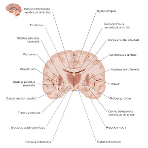
Coronal section of the brain (thalamus level): Gray matter structures (Latin)
Coronal section of the brain (thalamus level): Gray matter structures (Latin)
The most striking gray matter structure seen on this section are the thalami at the center of the illustration. Each thalamus is large, bilateral nucleus which faces its counterpart so that their medial surfaces comprise the lateral walls of the ventriculus tertius (third ventricle). Inferior to the thalamus is another nucleus called the nucleus subthalamicus and below that is the substantia nigra; a cluster of dopamine-releasing neurons in the mesencephalon. Lying laterally to these structures is the corpus striatum, often called the basal ganglia/nuclei basales. The corpus striatum is a collection of nuclei that consists of the nucleus caudatus (in the image only the truncus is visible), putamen and globus pallidus. The putamen and globus pallidus are often collectively referred to as the nucleus lentiformis due to their proximity and the lens-shape they create together. The last important gray matter structure is the hippocampus, which lies deep to the lobus temporalis.
Regular price
$7.56 USD
Regular price
Sale price
$7.56 USD
Unit price
per
Couldn't load pickup availability


#C87257
#B06C53
#6B2E16
#6C5A54
#E59A8A
#CEB5AD

