Liene Znotina
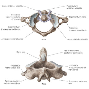
Cervical spine bones and ligaments: atlas and axis (Latin)
Cervical spine bones and ligaments: atlas and axis (Latin)
The atlas (C1) is a ring-shaped vertebra, devoid of a corpus vertebrae and processus spinosus. It consists of the anterior and posterior arcus vertebrae which are connected via a massa lateralis atlantis on each side, together enclosing the canalis vertebralis through which the spinal cord passes. Each massa lateralis atlantis features facies articulares on its superior and inferior surface and a single processus transversus that projects laterally.The anterior and posterior arcus vertebrae feature several bony landmarks that provide the attachment points for the ligaments of the columna vertebralis cervicalis. Moreover, the arcus vertebrae anterior contains a facies articularis that participates in formation of the art. atlantoaxialis mediana, a joint between atlas and axis. The facies articulares superior et inferior of the massae laterales atlantis participate in the artt. atlantooccipitales et atlantoaxiales lateralis, respectively. The former is a joint between the atlas and the os occipitale, while the latter is a three-part joint between the atlas and axis. The processus transversus that stem from the massae laterales atlantis feature a small foramen called foramen transversarium. This foramen exists in vertebrae C1-C6 and it is traversed by the a. vertebralis.The axis (C2) primarily differs from a typical vertebra by its prominent bony projection known as the dens axis (odontoid process). As a whole, the axis consists of anterior and posterior parts. The anterior part is formed by the corpus vertebrae and dens axis, while the posterior part consists of two pediculi, two processus transversus, two laminae and a processus spinosus. The corpus vertebrae of the axis is small and inferiorly elongated so that it overlaps the vertebra C3. It serves as an attachment point to several muscles and ligaments of the neck. The dens axis projects from the superior surface of the body. It features a facies articularis anterior dentis axis which articulates with atlas at the art. atlantoaxialis mediana and a facies articularis posterior dentis axis over which runs the lig. transversum atlantis. Either side of the dens axis, the corpus vertebrae of the axis features facies articulares superior et inferior which are located on its superior and inferior surfaces, respectively. The former participate in the artt. atlantoaxiales lateralis, while the latter articulate with vertebra C3 via specialized artt. zygapophysiales, which are sometimes collectively referred to as the art. vertebroaxialis.
Regular price
$7.56 USD
Regular price
Sale price
$7.56 USD
Unit price
per
Couldn't load pickup availability


