
Paul Kim
Cerebellum - Anterior view (Latin)
Cerebellum - Anterior view (Latin)
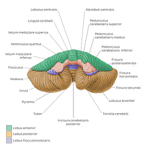
This view of the cerebellum is once again characterized by a central vermis which connects the hemispheria cerebelli. Also visible are three pairs of prominent fiber bundles, the pedunculi cerebellares superior, middle et inferior, that connect the cerebellum to the midbrain (mesencephalon), pons, and medulla oblongata, respectively. The vela medullares superior et inferior are thin sheets of white matter which form the tegmen ventriculi quarti. This is the only perspective in which the three lobi of the cerebellum (lobus anterior, lobus posterior and lobus flocculonodularis) are collectively visible. The facies anterior (petrosa) cerebelli bears the tonsillae cerebelli which protrude inferomedially between the tuber and uvula vermis. Each tonsilla is separated from the lobulus biventralis by the fissura secunda (a.k.a. fissura retrotonsillaris/postpyramidalis). Close to the center of the facies anterior cerebelli and anterior to the uvula vermis is the nodulus vermis; this connects with the two flocculi to form the lobus flocculonodularis. Each flocculus is located posterior to the pedunculus cerebellaris medius.
Regular price
$7.56 USD
Regular price
Sale price
$7.56 USD
Unit price
per
Couldn't load pickup availability

#C08F47
#645DA7
#64502A
#42392C
#E7C99E
#96CDB2