
Paul Kim
Cerebellum - Anterior view (English)
Cerebellum - Anterior view (English)
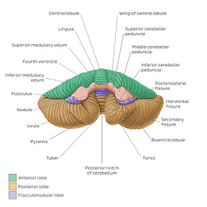
This view of the cerebellum is once again characterized by a central vermis which connects the cerebellar hemispheres. Also visible are three pairs of prominent fiber bundles, the superior, middle, and inferior cerebellar peduncles that connect the cerebellum to the midbrain, pons, and medulla oblongata, respectively. The superior and inferior medullary vela are thin sheets of white matter which form the roof of the fourth ventricle. This is the only perspective in which the three lobes of the cerebellum (anterior, posterior and flocculonodular) are collectively visible.The anterior (petrosal) surface of the cerebellum bears the tonsils of the cerebellum which protrude inferomedially between the tuber and uvula of the vermis. Each tonsil is separated from the biventral lobule by the secondary fissure (a.k.a. retrotonsillar/postpyramidal fissure). Close to the center of the anterior surface and anterior to the uvula is the nodule of vermis; this connects with the two flocculi to form the flocculonodular lobe. Each flocculus is located posterior to the middle cerebellar peduncle.
Regular price
$7.56 USD
Regular price
Sale price
$7.56 USD
Unit price
per
Couldn't load pickup availability

#C08F47
#645DA7
#64502A
#42392C
#E7C99D
#96CDB2