
Samantha Zimmerman
Calcaneus (Medial and lateral view) (Latin)
Calcaneus (Medial and lateral view) (Latin)
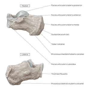
On the medial view, the processus anterior calcanei features the facies articularis cuboidea, which articulates with the os cuboideum to form the art. calcaneocuboidea. The facies articularis talaris anterior and facies articularis talaris media articulate with their calcaneal counterparts and contribute to the formation of the art. talocalcaneonavicularis of the tarsus. Posterior to the facies articularis talaris anterior is the aforementioned sustentaculum tali.Along the lateral surface of the calcaneus is a small prominence known as the trochlea fibularis. It is typically located between the tendons of the m. fibularis longus and m. fibularis brevis and serves as a second pulley for the fibularis tendons.
Regular price
$7.56 USD
Regular price
Sale price
$7.56 USD
Unit price
per
Couldn't load pickup availability

#897D73
#594738
#C5BCB4