
Samantha Zimmerman
Calcaneus (Medial and lateral view) (English)
Calcaneus (Medial and lateral view) (English)
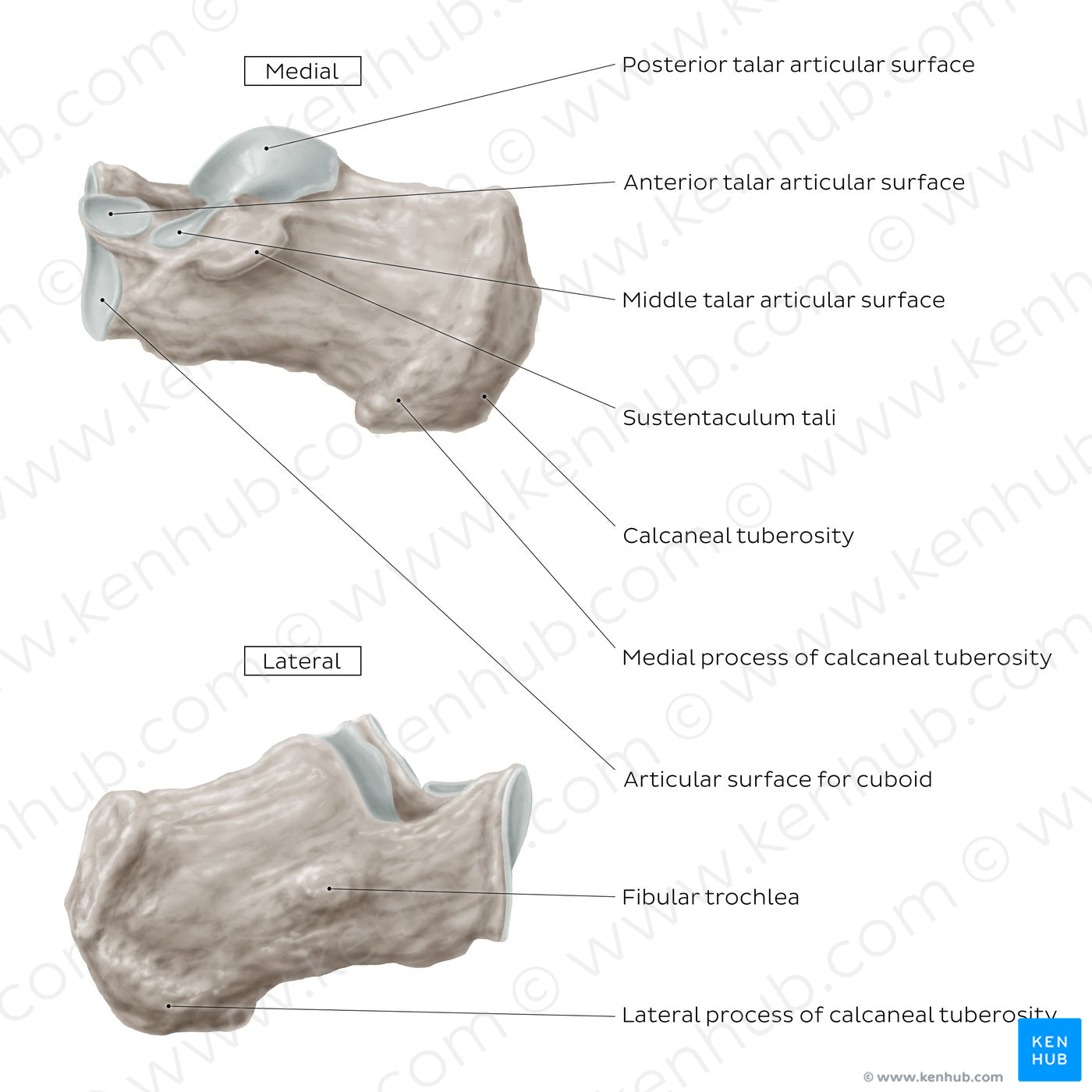
On the medial view, the anterior process of the calcaneus features the cuboid articular surface, which articulates with the cuboid bone to form the calcaneocuboid joint. The anterior and middle talar articular surfaces articulate with their calcaneal counterparts and contribute to the formation of the talocalcaneonavicular joint of the tarsus. Posterior to the anterior talar articular surface is the aforementioned sustentaculum tali.Along the lateral surface of the calcaneus is a small prominence known as the fibular trochlea. It is typically located between the tendons of the fibularis longus and brevis muscles and serves as a second pulley for the fibularis tendons.
Regular price
$7.56 USD
Regular price
Sale price
$7.56 USD
Unit price
per
Couldn't load pickup availability
#897D73
#594738
#C5BCB4