
Paul Kim
Bones of the orbit (Latin)
Bones of the orbit (Latin)
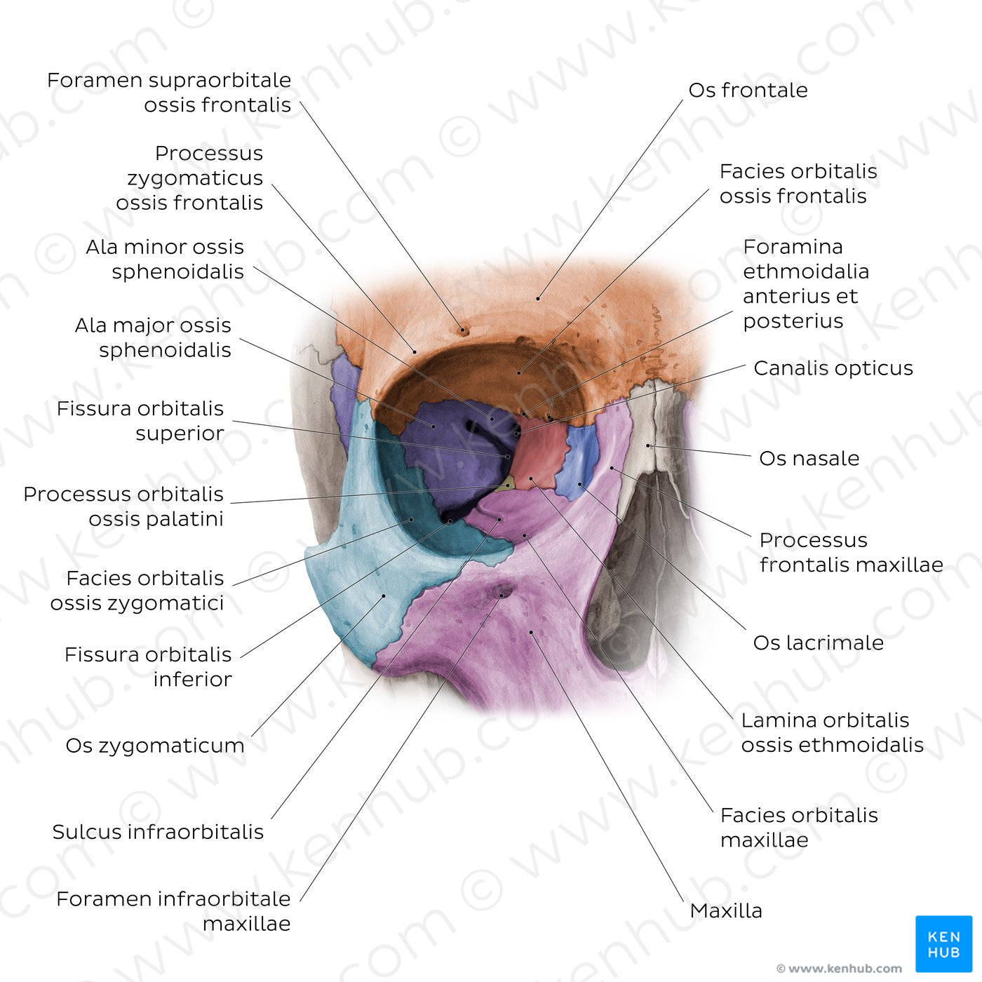
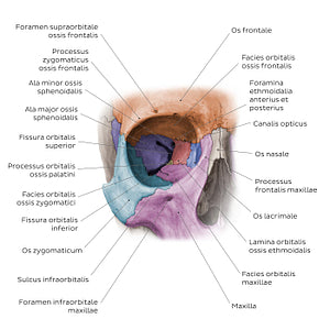
Anterior view of the orbita dextra showing its bony walls and associated fissures and foramina. The superior wall of the cavitas orbitae is formed mainly by the facies orbitalis ossis frontalis. The medial wall consists of the lamina orbitalis ossis ethmoidalis, processus frontalis maxillae, os lacrimale and os sphenoidale. The inferior wall of the orbit is formed by the facies orbitalis maxillae, processus orbitalis ossis palatini and os zygomaticum, while the lateral wall consists of the processus frontalis ossis zygomatici and the ala major ossis sphenoidalis.
Regular price
$7.56 USD
Regular price
Sale price
$7.56 USD
Unit price
per
Couldn't load pickup availability
#B9643E
#A26258
#693B25
#553728
#E2B197
#CDA2C4