
Paul Kim
Basal view of the brain (English)
Basal view of the brain (English)
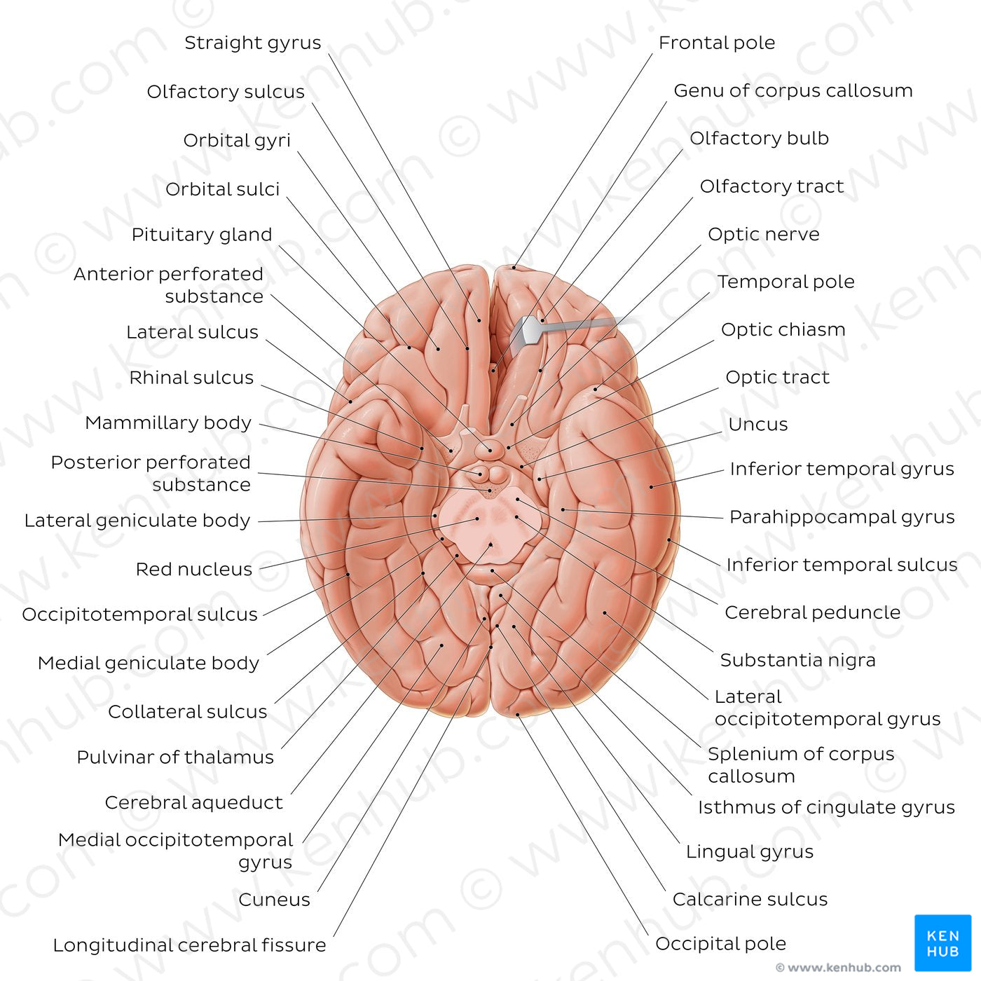
The inferior surface of the cerebrum is divided by the lateral sulcus into anterior and posterior parts. In the anterior part, the orbital gyri (anterior, posterior, medial and lateral orbital gyri) and straight gyrus can be identified. The orbital gyri are separated by the orbital sulci which together form an H shaped groove between the four gyri. The posterior part is marked by two primary anteroposteriorly oriented sulci: the collateral sulcus and occipitotemporal sulcus. These two sulci delineate the occipitotemporal gyri, which are divided by the midfusiform sulcus into medial and lateral occipitotemporal gyri. The lateral occipitotemporal gyrus is continuous with the inferior temporal gyrus around the inferolateral margin. The posterior part of the collateral sulcus runs parallel to the calcarine sulcus, and together they delineate the lingual gyrus. Besides the cortical structures, in this view the pituitary gland, the mammillary bodies and the geniculate bodies can also be appreciated. Lastly, a cross section of the brainstem on the midbrain level reveals its internal structure.
Regular price
$7.56 USD
Regular price
Sale price
$7.56 USD
Unit price
per
Couldn't load pickup availability
#D16B55
#94675A
#642E22
#6B5B54
#EE9E87
#D2B2A8