
Paul Kim
Abdominal surface of the diaphragm (Latin)
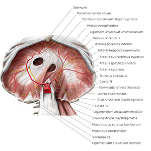
Abdominal surface of the diaphragm (Latin)
This inferior view of the abdominal surface of the diaphragma shows the muscular and tendinous components, as well as the three main openings of the diaphragm: the hiatus aorticus (aorta, v. azygos and ductus thoracicus), hiatus oesophagus (oesophagus, branches of a. et v. gastrica sinistra and truncus vagalis anterior) and foramen venae cavae (v. cava inferior and branches of the n. phrenicus dexter). These are formed with the help of tendinous structures, including the crus dextrum and sinistrum diaphragmatis as well as the lig. arcuatum medianum. Two further ligaments, the lig. arcuatum mediale and laterale, form openings posterior to the diaphragma, through which the m. psoas major and m. quadratus lumborum pass. Also visible in this inferior view of the diaphragm are components of the axial skeleton, such as the ribs, sternum and the first three vertebrae lumbales. The major vessels supplying the diaphragma are also seen travelling along its abdominal surface: the n. phrenicus and a. phrenica inferior.
Regular price
$7.56 USD
Regular price
Sale price
$7.56 USD
Unit price
per
Couldn't load pickup availability

#D41C27
#A65B62
#60211A
#594732
#E69B96
#D3B0AB