
Paul Kim
Surface anatomy of the brainstem (English)
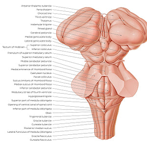
Surface anatomy of the brainstem (English)
The posterior part (tectum) of the midbrain has two pairs of raised, round protrusions that are collectively known as the quadrigeminal plate. This complex consists of the superior and inferior colliculi. Each inferior colliculus is separated from its contralateral counterpart by the frenulum of the superior medullary velum. The dorsal aspect of the pons and upper medulla oblongata forms the floor of the fourth ventricle, forming a large landmark known as the rhomboid fossa. The upper rhomboid fossa is divided in half by a median sulcus, with each half being further divided again by a parallel line, the sulcus limitans. Between the median sulcus and sulcus limitans is the medial eminence, while the region lateral to the sulcus limitans is the upper portion of the vestibular area. The caerulean nucleus/locus caeruleus is also found on the upper rhomboid fossa. The cranial/superior limit of the pons is formed by a structure which contributes to the formation of the roof of the fourth ventricle, the superior medullary velum, while the inferior boundary of the pons is formed by the medullary striae of the fourth ventricle. The dorsal/posterior surface of the medulla oblongata is divided into an open/superior part, which contains the caudal half of the fourth ventricle, and a closed/inferior part, which contains the central canal that continues into the spinal cord. Structures of the lower rhomboid fossa within the superior part of the medulla oblongata include the hypoglossal and vagal trigones, obex and the lower portion of the vestibular area. The dorsal aspect of the inferior part of the medulla oblongata is marked by a posterior median sulcus, which is bounded on either side by a raised structure known as the gracile tubercle which is in turn bordered laterally by the cuneate tubercle. The gracile and cuneate tubercles are continuations of the gracile and cuneate fasciculi of the spinal cord.
Precio habitual
$7.56 USD
Precio habitual
Precio de oferta
$7.56 USD
Precio unitario
por
No se pudo cargar la disponibilidad de retiro

#C4623B
#94655A
#692720
#726E6C
#F2AB97 y #D1B3A9